| Bitewing | ● Examine the crowns of the upper and lower teeth in one section of the mouth. ● Identify cavities development in hard-to-reach areas such as in between the teeth, underneath old fillings or crowns. |
| Periapical | ● Display a tooth from the crown to the root in order to examine one or two teeth for root problems, cavities, and bone loss due to periodontal diseases |
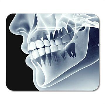
| Panoramic | ● Show the entire mouth. ● Detect periodontal disease, infections, jaw fractures, cysts and tumors. ● Track tooth development in pediatric patients and assess the need for orthodontic intervention. ● These images are also helpful in evaluating the need for wisdom teeth removal. |
| Occlusal | ● Show the arch of the teeth from the top or the bottom jaw. ● Used to detect pathologies such as clefts, cysts or extra teeth. |
| Cone Beam Computerized Tomography (CBCT) | ● Precise 3D view of the patient’s mouth. ● Helpful in assessing the development and space of their teeth and planning the placement of dental implants. |
Much like how you can’t see through walls, we can’t see through teeth. For example, without X-rays, your dentist will not be able to detect decay between the teeth or underneath a filling. Without up-to-date radiographs once a year, we are leaving areas of your mouth unchecked, allowing potential issues to worsen quickly. Cavities do not always hurt and it is best to treat them before they lead to infections and possible tooth loss. From early caries detection to early gum disease, there are numerous oral issues that can be caught early enough to reverse or treat before they turn into bigger problems. The earlier we catch a problem, the lower the cost will be to correct it.
Many factors such as age, history, the current state of your medical and oral health, and presence of gingivitis or periodontitis can influence how often dental X-rays are needed. Patients who may need X-rays taken more often include: children and teenagers who have a history of many cavities, adults with many restorations (fillings, crowns, bridges), patients with periodontal disease and dry mouth, and smokers and tobacco chewers.
Are dental X-ray safe? We are exposed to natural sources of radiation (cosmic rays, radon emission) all the time. On average, Americans receive about 300 millirem of natural radiation annually according to the U.S. Nuclear Regulatory Commission. The use of digital X-rays reduces radiation by as much as 80% compared to X-ray radiography. According to the American Dental Association (ADA), radiation exposure due to dental X-rays is minimal in comparison to both human-made and natural radiation sources. For example, the amount of radiation from dental X-ray is 0.5 millirem, in comparison to 10 millirem from one adult chest X-ray. Dental radiographs account for about 2.5% of the radiation dose received from all medical X-ray imaging procedures.
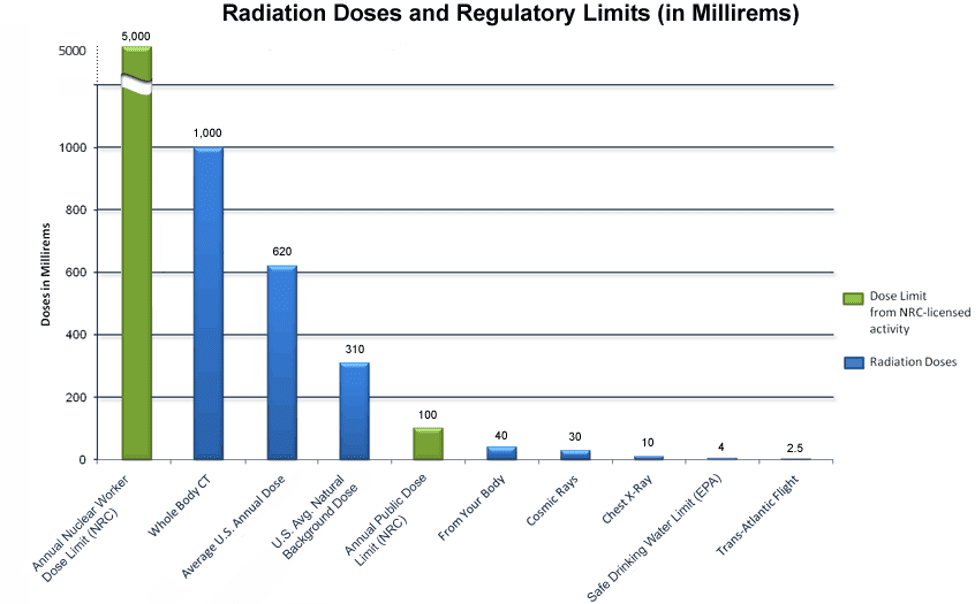
Source: nrc.gov
| DENTAL | Procedure | Approximate effective radiation dose | Comparable to natural background radiation for: |
|---|---|---|---|
 |
Dental X-ray | 0.5 millirem | 1 day |
| Panoramic X-ray | 2.5 millirem | 3 days |
Source: https://www.radiologyinfo.org/en/info/safety-xray